What Can You See With Foldscope 140x Magnification Cell Examples?
A foldscope with 140x magnification opens a microscopic world of cellular detail that rivals traditional lab microscopes. At this magnification level, you can observe individual cells, organelles, and microbial structures with stunning clarity. The foldscope’s affordable paper design makes this technology accessible to students, educators, and citizen scientists worldwide. In June 2026, thousands of classrooms use foldscopes to explore real biological specimens. This guide reveals exactly what cell examples you can examine and how to prepare them for optimal viewing.
The 140x magnification rating represents a significant leap in optical capability. You’ll move beyond simple tissue observation into the realm of subcellular structures. Plant cells become transparent windows into chloroplasts and cell walls. Animal cells reveal nuclei and mitochondrial patterns. Microorganisms like bacteria and protists become clearly visible subjects. The foldscope achieves this through a precisely engineered spherical lens system mounted in a folded paper body.
Key Takeaway: Foldscope 140x magnification provides sufficient optical power to observe detailed cellular structures, making it ideal for educational exploration of biological specimens.
Which Cell Types Show Best Results at 140x Magnification?
Certain cell types display remarkable detail under 140x magnification. Plant cells emerge as the most visually impressive specimens due to their rigid cell walls and large organelles. Onion epidermal cells reveal distinct cell boundaries and nuclei. Elodea leaf cells showcase chloroplasts in vibrant green patterns. Animal cells from cheek swabs display nuclei prominently against cytoplasmic backgrounds. Microorganisms including paramecia, euglena, and various bacteria become identifiable subjects.
The key to successful observation lies in specimen selection and preparation. Cells with natural pigmentation—like plant tissues containing chlorophyll—appear more dramatic. Transparent specimens work best because light passes through easily. Larger cells, such as plant cells and protists, show more internal detail than tiny bacteria. Staining enhances visibility of structures that lack natural color.
Plant Cell Examples at 140x Magnification
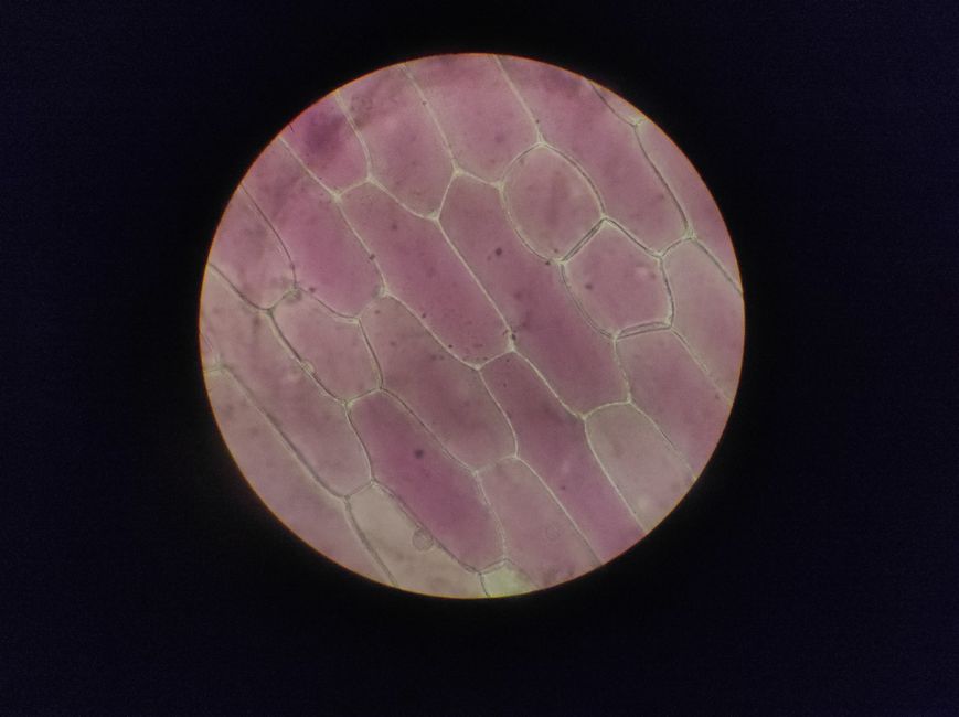
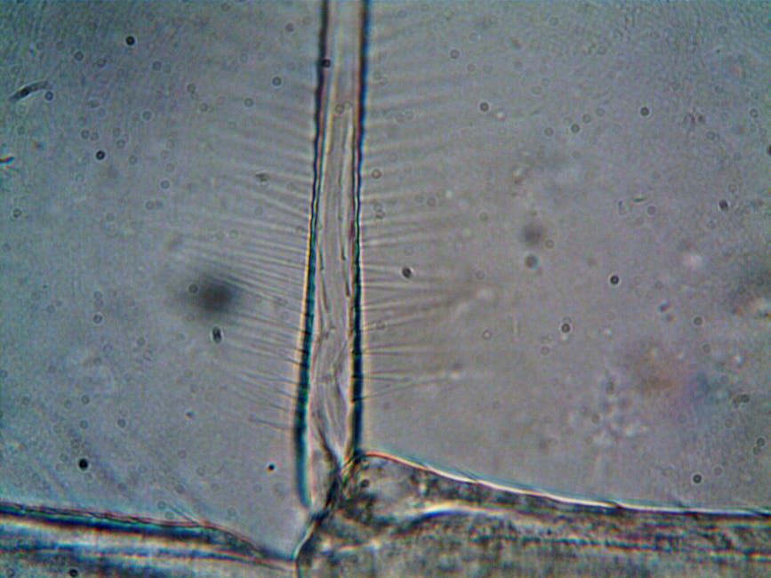
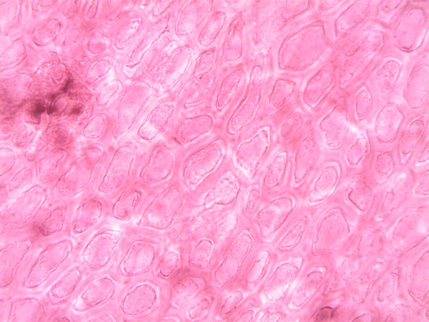
Plant cells deliver the most impressive visual results under foldscope observation. The rigid cell wall creates a clear rectangular boundary around each cell. Chloroplasts appear as bright green organelles distributed throughout the cytoplasm. The nucleus sits centrally, often visible as a darker circular structure. Vacuoles occupy significant space, creating a distinctive cellular architecture.
- Onion epidermal cells show clear cell walls and nuclei without staining.
- Elodea leaf cells display vibrant chloroplasts in organized patterns.
- Tomato skin cells reveal pigmented structures and detailed wall patterns.
- Moss leaf cells show distinctive cell wall arrangements and organelle distribution.
Animal Cell Examples at 140x Magnification
Animal cells present different challenges but equally rewarding observations. Without cell walls, animal cells appear more rounded and less defined than plant cells. The nucleus becomes the dominant visible structure, appearing as a dark central sphere. Cytoplasm fills the remaining space, sometimes showing granular texture. Cheek cells, blood cells, and nerve cells each reveal distinctive characteristics.
- Cheek epithelial cells show large nuclei and clear cell membranes.
- Red blood cells appear as pale, flat discs lacking nuclei.
- White blood cells display irregular shapes and visible nuclei.
- Nerve cells reveal extended axons and dendrites when properly prepared.
Key Takeaway: Plant cells offer superior visual results due to cell walls and chloroplasts, while animal cells require careful staining but reveal important nuclear structures.
How Do You Prepare Specimens for Optimal 140x Magnification Viewing?
Proper specimen preparation dramatically improves observation quality at 140x magnification. The foldscope’s shallow depth of field demands thin, transparent samples. Mounting medium selection affects image clarity and specimen longevity. Staining techniques highlight structures invisible under bright-field illumination. Temperature and humidity also influence cellular appearance and specimen preservation.
Essential Preparation Techniques
Begin with fresh, living specimens whenever possible. Living cells display natural movement and color that fixed specimens cannot match. Create thin smears on glass slides using gentle spreading motions. Allow smears to air-dry completely before adding mounting medium. This prevents air bubbles that scatter light and degrade image quality.
- Collect plant tissue from healthy, disease-free specimens for best results.
- Prepare cheek cells by gently scraping the inside of your mouth with a clean toothpick.
- Use distilled water as initial mounting medium for living specimens.
- Apply cover slips carefully to avoid trapping air bubbles under the sample.
- Wait 2-3 minutes before viewing to allow specimens to settle and flatten.
Staining Methods for Enhanced Visibility
Staining transforms nearly invisible structures into vivid, observable features. Methylene blue highlights nuclei and certain organelles with deep blue coloration. Iodine stains starch granules in plant cells, creating dramatic contrast. Crystal violet penetrates cell membranes and stains nuclei intensely. Each stain requires specific preparation protocols and observation timing.
Apply stains carefully using a dropper at the slide edge. Allow the stain to diffuse gradually through the specimen rather than flooding the entire slide. Excess stain obscures detail and creates background noise. Wait 30-60 seconds for full staining before observation. Rinse excess stain with distilled water if the image appears too dark.
Key Takeaway: Thin, properly mounted specimens with appropriate staining reveal cellular structures clearly at 140x magnification, requiring patient preparation techniques.
What Magnification Limitations Should You Understand?
The 140x magnification level represents a sweet spot between affordability and optical capability. However, important limitations exist that affect what you can observe. Subcellular structures like ribosomes and individual thylakoids remain invisible. Virus particles and most bacteria appear as fuzzy dots rather than distinct organisms. The foldscope’s shallow depth of field means only a thin optical section appears in focus simultaneously.
Resolution—the ability to distinguish two separate points—decreases as magnification increases. At 140x, the theoretical resolution limit approaches 0.5 micrometers under ideal conditions. Practical resolution often reaches only 1-2 micrometers due to optical imperfections and lighting challenges. This means organelles smaller than one micrometer blur together into indistinct masses.
- Ribosomes (25 nanometers) remain completely invisible at this magnification level.
- Mitochondrial cristae structure appears as blurry internal patterns.
- Bacterial flagella appear as faint lines rather than distinct structures.
- Virus particles require electron microscopy for direct observation.
Key Takeaway: Foldscope 140x magnification excels at cellular-level observation but cannot resolve subcellular organelles or microorganisms below one micrometer.
How Does Foldscope 140x Compare to Traditional Microscopes?
Traditional compound microscopes typically offer 40x to 1000x magnification ranges. The foldscope’s 140x magnification falls in the lower-middle range of traditional microscope capability. However, cost differences are dramatic—a foldscope costs under twenty dollars while quality compound microscopes exceed five hundred dollars. The foldscope prioritizes accessibility and ease of use over maximum magnification power.
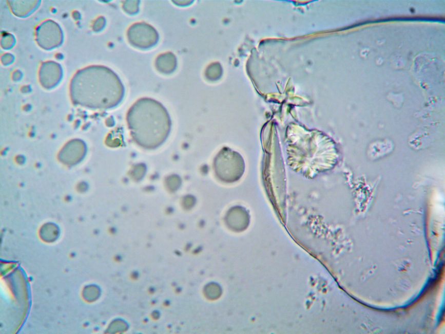
Image quality comparisons reveal interesting trade-offs. Traditional microscopes provide brighter, clearer images due to superior optics and powerful illumination systems. Foldscopes depend on ambient light or simple LED illumination, creating dimmer fields of view. However, foldscopes excel in portability and durability. A foldscope fits in a pocket and survives drops that would destroy a traditional microscope.
Practical Advantages of Foldscope 140x
- Cost: Under twenty dollars versus hundreds for traditional microscopes.
- Portability: Fits in a pocket or backpack for field observations.
- Durability: Paper construction survives drops and rough handling.
- Ease of use: Simple assembly and intuitive operation for all ages.
- Accessibility: Enables microscopy education in resource-limited regions.
For serious scientific work, traditional microscopes remain superior. For educational exploration, citizen science, and field investigations, the foldscope offers unmatched value. Many educators use foldscopes as entry points before transitioning students to traditional lab microscopes. The 140x magnification level provides sufficient detail for most high school biology curricula.
Key Takeaway: Foldscope 140x magnification cannot match traditional microscope image quality, but offers superior cost, portability, and accessibility for educational use.
What Practical Tips Improve Your Foldscope 140x Observations?
Mastering foldscope observation requires developing specific techniques and practices. Lighting conditions dramatically affect image quality and visibility. Focus precision demands patience and gentle hand movements. Specimen preparation quality determines whether you observe cellular details or blurry masses. These practical tips transform casual observations into productive scientific investigations.
Lighting and Focus Techniques
- Use natural daylight or bright LED illumination for clearest images.
- Position light sources at 45-degree angles to minimize glare and shadows.
- Focus slowly by turning the focus wheel in small increments.
- Rock the specimen slightly to find the optimal focal plane.
- Allow your eyes to adjust to the dimmer field of view for 30-60 seconds.
- Avoid direct sunlight that creates excessive brightness and heat.
Specimen Handling Best Practices
Handle specimens gently to prevent damage and maintain observation quality. Glass slides are fragile and require careful storage. Keep prepared slides in protective cases away from dust and moisture. Label slides with specimen type and preparation date for future reference. Store slides in cool, dark locations to preserve stains and prevent fading.
Fresh specimens often yield superior observations compared to preserved materials. Collect specimens shortly before observation when possible. Keep living specimens moist until observation to maintain cellular integrity. Avoid exposing specimens to extreme temperatures that cause cellular damage. Document your observations through sketches or photographs for later analysis.
Common Mistakes to Avoid
- Using thick specimens that don’t fit within the shallow depth of field.
- Applying excessive stain that obscures rather than highlights structures.
- Focusing too quickly and missing the optimal focal plane.
- Touching the lens with fingers, which leaves smudges and reduces clarity.
- Attempting to observe specimens without proper mounting medium.
Key Takeaway: Proper lighting, careful focusing, and meticulous specimen preparation transform foldscope observations from disappointing to scientifically valuable.
Which Resources Support Foldscope 140x Learning and Observation?
The foldscope community offers extensive resources for users seeking to maximize their observations. Online databases catalog specimen preparation protocols and expected results. Educational institutions have adopted foldscopes as primary microscopy tools. Research initiatives use foldscopes for large-scale environmental monitoring projects. These resources democratize microscopy education and scientific investigation.
The Foldscope Imaging Project provides a global platform for sharing observations and discoveries. Teachers access curriculum materials aligned with national science standards. Student communities document their findings and compare observations across regions. This collaborative approach transforms individual observations into collective scientific knowledge. For comprehensive guidance on educational applications, explore the Foldscope 2.0 Explorer Kit for High School: Complete Guide June 2026.
- Foldscope.com provides official documentation and specimen protocols.
- YouTube channels demonstrate preparation techniques and observation methods.
- Research publications document foldscope applications in malaria diagnosis and environmental monitoring.
- Educational workshops teach microscopy fundamentals using foldscope technology.
Key Takeaway: Extensive online resources and educational communities support foldscope users in developing observation skills and sharing discoveries globally.
Can You Identify Specific Microorganisms at 140x Magnification?
Microorganism identification becomes possible at 140x magnification for larger species. Paramecia, with lengths of 50-300 micrometers, appear as distinct organisms with visible internal structures. Euglena displays characteristic flagella and eyespots. Amoebas reveal pseudopods and nuclei clearly. Algae species show distinctive cell arrangements and pigmentation patterns. However, bacteria remain challenging subjects due to their tiny size.
Bacteria appear as fuzzy dots or short rods at 140x magnification. Individual bacterial cells measure 0.5-5 micrometers, pushing the limits of foldscope resolution. Bacterial colonies become visible, but individual organisms blur together. Cyanobacteria and filamentous bacteria show better results due to larger size and distinctive arrangements. Staining becomes essential for any bacterial observation.
Observable Microorganisms at 140x
- Paramecia: Visible as slipper-shaped organisms with internal ciliary patterns.
- Euglena: Green flagellates showing characteristic eyespots and flagella.
- Amoebas: Shapeshifting organisms with visible pseudopods and nuclei.
- Algae: Green or brown cells showing chloroplasts and cell wall structures.
- Cyanobacteria: Blue-green filaments visible as organized colonies.
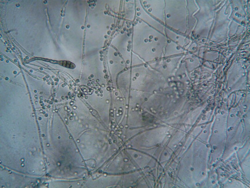
- Mold spores: Distinctive structures at tips of fungal hyphae.
Key Takeaway: Larger microorganisms like paramecia and euglena appear clearly at 140x magnification, while bacteria remain at the resolution limit and require staining.
What Are the Best Cell Examples for Beginning Foldscope Users?
Beginners achieve best results with forgiving specimens that display clear structures. Onion epidermal cells remain the gold standard starting specimen. These cells feature obvious cell walls, nuclei, and vacuoles without requiring stains. Elodea leaves provide equally impressive results with vibrant chloroplasts. Both specimens are readily available and require minimal preparation time.
Cheek cells offer accessible animal cell examples for all ages. A simple toothpick scrape provides sufficient material for multiple slides. The nucleus appears prominently without staining, though methylene blue enhances visibility. Water fleas (Daphnia) work wonderfully for observing living organisms in motion. Their transparent bodies reveal internal structures and movement patterns clearly.
Beginner-Friendly Cell Specimens
- Onion epidermal cells: No staining required, clear cell walls and nuclei visible.
- Elodea leaf cells: Natural green color highlights chloroplasts beautifully.
- Cheek epithelial cells: Easy collection, large nuclei, minimal preparation needed.
- Water fleas: Living organisms showing internal organs and movement.
- Lettuce leaf cells: Similar to onion but larger, showing excellent detail.
- Moss leaf cells: Distinctive arrangements with visible chloroplasts and walls.
Start with these accessible specimens before attempting challenging preparations. Success builds confidence and motivates continued exploration. Once comfortable with basic techniques, progress to stained specimens and microorganisms. This graduated approach develops observation skills systematically.
Key Takeaway: Onion and Elodea cells provide ideal starting points for beginners, requiring minimal preparation while displaying impressive cellular detail.
How Has Foldscope Technology Evolved for Better 140x Observations?
Since its introduction in 2014, foldscope technology has undergone continuous refinement. Early versions struggled with optical aberrations and inconsistent magnification. Current 140x models feature improved lens quality and more precise assembly tolerances. Manufacturing processes have become more rigorous, ensuring consistent performance across units. These improvements translate directly into clearer, brighter observations.
The foldscope community drives innovation through feedback and collaborative development. Researchers identified optimal lens materials and geometries through extensive testing. Assembly instructions have been simplified, reducing manufacturing errors. LED illumination options have been added, improving observation in low-light conditions. The 2026 generation of foldscopes represents the culmination of over a decade of refinement.
- Improved lens coatings reduce glare and increase light transmission.
- Better-quality paper stock provides more stable optical geometry.
- Refined focus mechanisms enable more precise focal adjustments.
- Optional LED illuminators extend observation possibilities beyond daylight.
- Standardized assembly processes ensure consistent optical performance.
Key Takeaway: Modern foldscope 140x models benefit from over a decade of technological refinement, delivering superior optical quality compared to earlier generations.
What Scientific Projects Use Foldscope 140x Magnification?
Beyond classroom education, foldscopes enable genuine scientific research. Malaria diagnosis programs in Africa use foldscopes to detect parasites in blood samples. Environmental monitoring projects track water quality by observing microorganism populations. Biodiversity surveys document microscopic organisms in soil and water samples. Citizen science initiatives engage thousands of participants in data collection and analysis.
The affordability and portability of foldscopes make them ideal for field research in resource-limited regions. Researchers can deploy hundreds of units simultaneously, collecting data at unprecedented scales. Data collected through foldscope observations contributes to peer-reviewed publications and policy decisions. This democratization of microscopy enables scientific advancement in underserved communities.
- Malaria diagnosis: Detecting Plasmodium parasites in blood smears accurately.
- Water quality monitoring: Observing indicator organisms in environmental samples.
- Soil biodiversity: Cataloging microorganisms and small invertebrates in ecosystems.
- Disease surveillance: Tracking parasite prevalence in public health programs.
- Educational research: Studying learning outcomes when using foldscopes.
Key Takeaway: Foldscope 140x magnification enables meaningful scientific research and public health applications, particularly in resource-limited regions worldwide.
What Final Insights Should You Remember About Foldscope 140x Magnification?
Foldscope 140x magnification represents a remarkable achievement in accessible microscopy. This affordable paper device opens the microscopic world to students, educators, and researchers worldwide. While limitations exist compared to traditional microscopes, the foldscope’s unique advantages make it invaluable for education and field research. Proper specimen preparation and careful observation techniques unlock the full potential of this technology.
The cell examples visible at 140x magnification span the entire spectrum of biology. Plant cells showcase chloroplasts and cell walls with stunning clarity. Animal cells reveal nuclei and cytoplasmic structures. Microorganisms from paramecia to algae become identifiable subjects. Bacteria push the resolution limits but remain observable with appropriate staining. This diversity of observable structures makes foldscope exploration endlessly rewarding.
As you develop your foldscope skills, remember that observation is both art and science. Patience, careful preparation, and attention to lighting conditions transform casual observations into discoveries. Join the global foldscope community to share your findings and learn from others. Whether you’re a student exploring biology or a researcher advancing science, foldscope 140x magnification opens doors to understanding the microscopic world.
Key Takeaway: Foldscope 140x magnification democratizes microscopy, enabling meaningful biological observation and scientific discovery for anyone, anywhere, at minimal cost.
Frequently Asked Questions
Can you see bacteria clearly with foldscope 140x magnification?
Bacteria appear as small dots or rods at 140x magnification, pushing the resolution limits of foldscope technology. Individual bacterial cells measure 0.5-5 micrometers, making them barely visible. Staining techniques significantly improve bacterial visibility by adding contrast. Larger bacteria and bacterial colonies show better results than isolated cells. For clear bacterial observation, traditional microscopes at 1000x magnification work better.
How long does it take to see cellular detail in a foldscope?
After proper specimen preparation and mounting, cellular detail becomes visible within seconds. However, achieving optimal focus and lighting may require 2-5 minutes of adjustment. Your eyes need 30-60 seconds to adjust to the dim field of view. Specimen preparation itself takes 5-15 minutes depending on complexity. Total time from specimen collection to observation ranges from 10-30 minutes.
Do you need stains to observe cells with foldscope 140x?
Staining is not absolutely required but dramatically improves visibility of many structures. Plant cells with chlorophyll appear vibrant without stains. Animal cells benefit enormously from methylene blue or crystal violet staining. Bacteria remain nearly invisible without staining. Living organisms display natural movement without stains, revealing behavioral characteristics. Start without stains, then experiment with staining as your skills develop.
What is the actual resolution limit of foldscope 140x magnification?
The theoretical resolution limit approaches 0.5 micrometers under ideal conditions. Practical resolution typically reaches 1-2 micrometers due to optical imperfections and lighting limitations. This means structures smaller than one micrometer blur together indistinctly. Organelles like mitochondria appear as fuzzy masses rather than distinct structures. Ribosomes and membrane proteins remain completely invisible at this magnification.
Can foldscope 140x observe living cells in real-time?
Yes, foldscope 140x can observe living cells and organisms in real-time. Paramecia, water fleas, and algae display movement and behavior clearly. Living plant cells show cytoplasmic streaming and organelle movement. However, the shallow depth of field makes tracking moving organisms challenging. Mount living specimens in water or appropriate culture media to maintain viability during observation.
How does light quality affect foldscope 140x observations?
Light quality dramatically impacts image brightness, contrast, and clarity. Natural daylight provides excellent illumination but varies with time and weather. Bright LED lights offer consistent, adjustable illumination. Avoid direct sunlight that creates excessive heat and glare. Diffused light from north-facing windows provides ideal conditions. Poor lighting results in dim, low-contrast images that obscure cellular detail.


Write Your Review
No reviews yet. Be the first to share your experience!